NEURODOME was created by an interdisciplinary team of scientists, digital effects artists, and planetarium production specialists who are harnessing revolutionary brain-imaging techniques to provide a glimpse into the inner workings of the mind. Producing and visualizing new scientific data is what we do. We’ll show you real images, not simulations, straight from the scientists themselves.
Meet the Data
Real images, real scientists.
Data Scientists
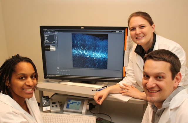
Dr. Ashlan Reid, Hassana Oyibo, & Martin Davis
Cold Spring Harbor Laboratory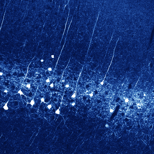
In their words:
Neurons in different brain regions perform different functions and must communicate with each other for the brain to process information and control behavior. Neurons communicate across long distances by sending axons far away from their cell bodies. We are interested in discovering which neurons communicate with particular brain regions so we designed a technique identify them.
We used a type of mouse where cells fluoresce red in the presence of a protein called cre recombinase. Since cre is not a native protein in the brain, none of the neurons glow until we add it. We built a genetically altered virus to deliver the gene for cre to particular neurons by infecting their axons. By making a small virus injection into one brain area, we can light up the cell bodies of neurons communicating with that area from far away.
Timothy Machado
Columbia University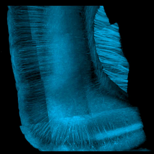
In his words:
We used a laser scanning microscope to take images of neurons deep in the brain. These neurons were genetically modified to express a fluorescent protein that glows only when you excite it with a laser. By scanning the laser across the whole 3D volume of the brain tissue, we could generate 3D images of almost all of the neurons present in our tissue sample. This technique lets us look at how different genes are expressed in different kinds of neurons in the brain. Understanding how many different kinds of neurons there are in the brain is important, because each type of neuron might have a unique role in processing information.

Dr. Sally A. Marik
The Rockefeller University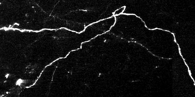
In her words:
In the cerebral cortex, millions of neurons integrate information from the five senses to create the organism’s perception of the world. Every neuron makes connections via their axons with thousands of other neurons across multiple locations within the cortex and throughout the brain. Mapping out exactly how these connections are maintained and altered in the adult is crucial to our understanding of the brain and its functions and dysfunctions, such as memory formation and
neurodegenerative diseases.I visualize axons within the cortex by using a virus that delivers GFP, a green fluorescent protein that was first extracted from jellyfish. The labeled neurons and their axons can be observed with a two-photon microscope. Axons are visualized, recorded, and compared over time. Under normal conditions, axonal structure is stable, but new experiences (such as learning, trauma, or disease) alter the connections or the entire structure by creating completely new axons and removing already existing ones. This process of axon remodeling may be a key to learning and memory formation.
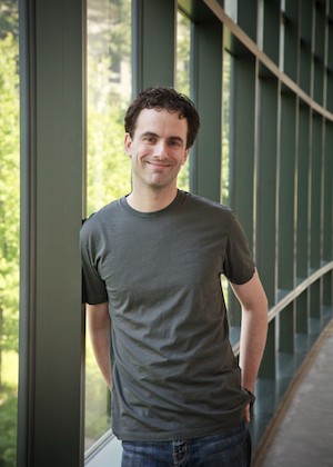
Gerald Sun
Johns Hopkins University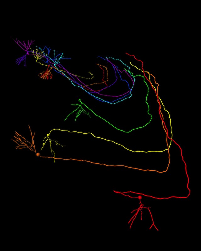
In his words:
"We developed a technique to capture images of neurons in their entirety in very large 3D volumes of brain tissue. We then used powerful computers and 3D glasses to visualize and trace the fine, complex structures of these neurons as shown here. Our data help provide a blueprint for some of the types of complex wiring that exist in the brain."
Gerry studies neural stem cells and newborn neuron development as a Neuroscience PhD student at Johns Hopkins University. This interest has led him to imaging fine structure and detail of stem cells and neurons and his images have been featured on the cover of The Journal of Neuroscience. His work has been supported in part by the Children's Tumor Foundation.
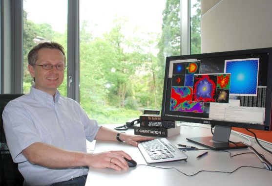
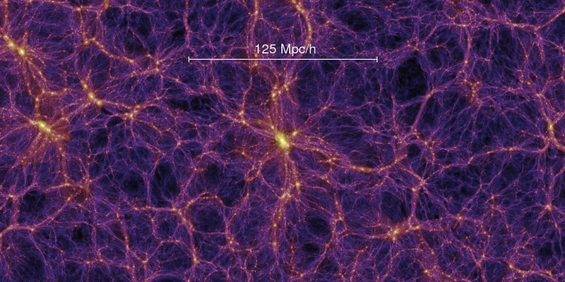
Dr. Volker Springel
University of Heidelberg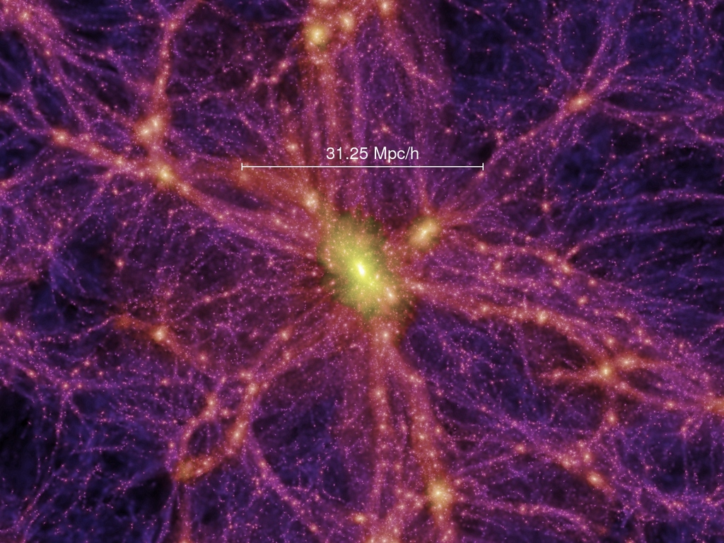
In his words:
The data showcased here are part of the "Millennium Simulation," a computer model that employed more than 10 billion particles of matter to trace the evolution of a 2 billion light-year cubic region of the Universe; the largest ever model of its kind at the time (Springel et. al, 2005). Simulations such as these can reveal the physical processes underlying the build-up of real galaxies and black holes.
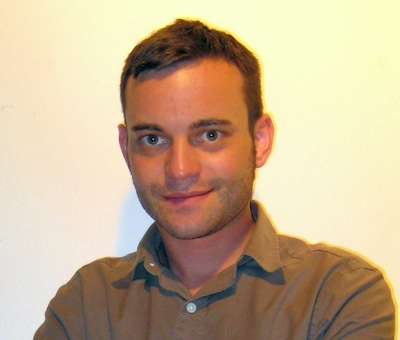
Dr. Joseph P. Zinter
Center for Engineering Innovation & Design, Yale University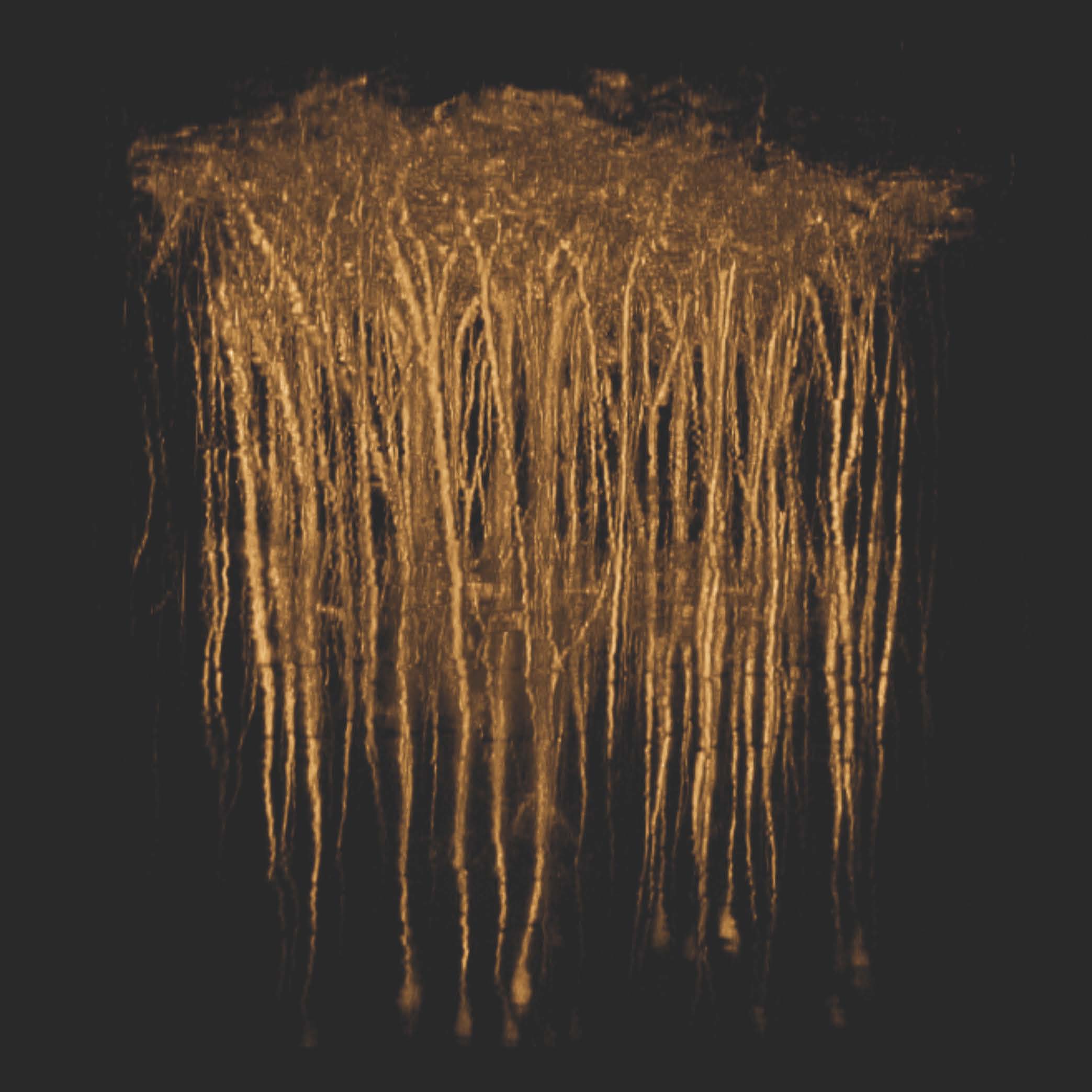
In their words:
We optimize microscope designs to image neurons deep in the brain of living animals using nonlinear laser scanning microscopy. This image stack was taken in the cortex of a mouse expressing a yellow fluorescence protein in neurons located almost 1-mm below the surface of cortex. Incidentally, we designed, fabricated, and built the scope in house!